Electron Microscopy
Connectomics
Mapping every synapse / Mouse visual cortex · MICrONS · NIH BRAIN CONNECTS
A wiring diagram of the cortex,
synapse by synapse.
The EM Connectomics project at the Allen Institute applies high-throughput serial-section electron microscopy to reconstruct neural circuits at synaptic resolution — capturing every cell, every process, and every connection within large volumes of brain tissue.
Work has focused primarily on mouse visual cortex, producing the largest functional connectomics datasets ever assembled. Reconstructions are paired with calcium imaging data from the same neurons, linking circuit structure directly to function.
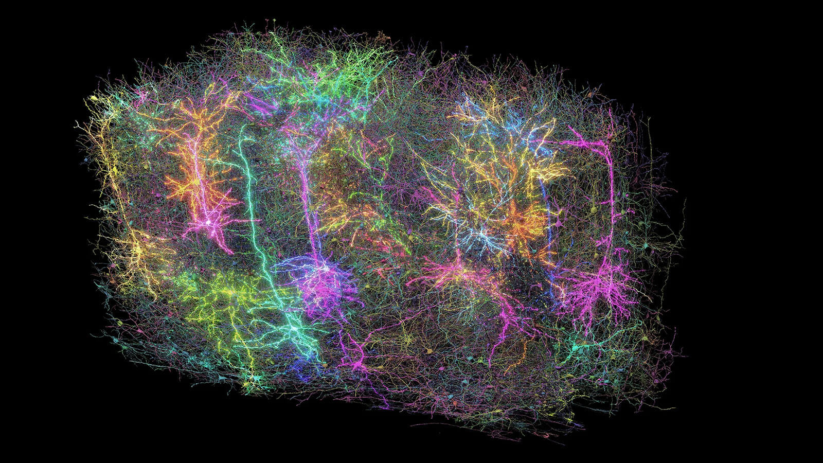
Neural circuit reconstruction from mouse visual cortex. Allen Institute for Brain Science / microns-explorer.org
The team combines expertise across electron microscopy, neuroscience, computer science, and mechanical engineering. Automated segmentation pipelines process petascale image volumes, and all resulting data are released openly to the scientific community through the MICrONS Explorer portal.
Machine Intelligence from
Cortical Networks.
The MICrONS program — funded by IARPA and led by a consortium at the Allen Institute, Princeton University, and Baylor College of Medicine — sought to reverse-engineer the algorithms of the brain to advance machine learning. It produced the most complete functional connectomics map of any cortical volume to date: a cubic millimeter of mouse visual cortex with dense calcium imaging co-registered to EM reconstruction of the same neurons.


Key findings from the dataset include the discovery that neurons with similar visual response properties preferentially form synaptic connections — a "like-to-like" wiring rule that also emerges independently in artificial neural networks trained on visual tasks.
Explorer
Explore the data
The full cubic millimeter dataset — including EM reconstructions, functional imaging, synaptic connectivity tables, and interactive Neuroglancer views — is freely available to the research community.
Open microns-explorer.org →Selected papers from
the MICrONS datasets.
Functional connectomics spanning multiple areas of mouse visual cortex
Dense calcium imaging combined with co-registered high-resolution EM reconstruction provides a functional connectomics map of tens of thousands of neurons across primary cortex and higher visual areas.
DOI 10.1038/s41586-025-08790-w →The synaptic architecture of layer 5 thick tufted excitatory neurons in mouse visual cortex
Maps the connections of layer 5 pyramidal neurons, revealing distinct local and intercortical wiring patterns, with an open framework for exploring cell-type connectivity.
DOI 10.1038/s41593-025-02004-2 →Inhibitory specificity from a connectomic census of mouse visual cortex
Uses volumetric EM to map and analyze the structure of cortical inhibition with synaptic resolution across a column of visual cortex.
DOI 10.1038/s41586-024-07780-8 →Functional connectomics reveals general wiring rule in mouse visual cortex
Neurons with similar response properties preferentially connect — a like-to-like rule that emerges within and across brain areas and layers, and independently arises in artificial neural networks.
DOI 10.1038/s41586-025-08840-3 →Allen Institute
Connectomics.
The Connectomics department — led by Nuno da Costa, Forrest Collman, and Clay Reid — is part of the Allen Institute's Brain Science division. The department's full scope, team, and ongoing projects are described on the Allen Institute website.
Institute
Connectomics at the Allen Institute
Full department overview, team members, EM and axonal connectomics projects, and links to open datasets.
Visit alleninstitute.org →