Clay Reid
EM Connectomics →Mapping the wired brain / Axonal connectomics · Human white matter · EM circuits
Tracing individual axons
through the human brain.
The Reid lab has launched a new research program to map long-range projection axons through human white matter at the scale of centimeters — something never achieved before. Using post-mortem human tissue, dense antibody staining, tissue expansion, and light-sheet fluorescence microscopy, the lab is characterizing the 3D trajectories and architectural organization of individual myelinated axons across regions of the entire brain.
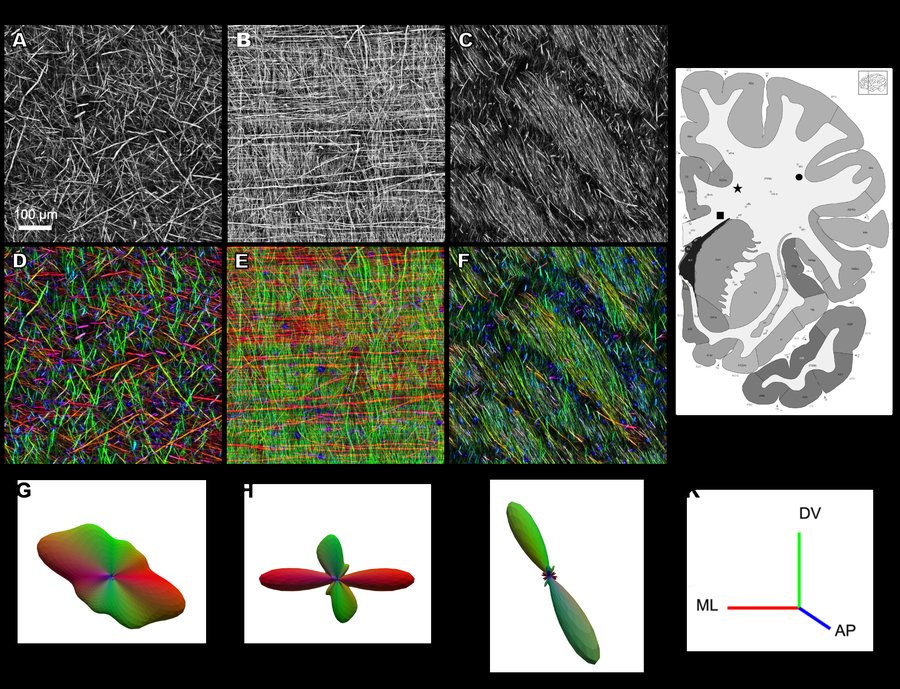
Heterogeneity of white-matter organization in the human brain
White matter makes up nearly half the human brain, yet the 3D organization of individual axons has never been directly characterized. Using a histological pipeline optimized for post-mortem human tissue, this work reveals striking regional diversity — from loosely packed meshworks in superficial white matter to tightly packed parallel bundles in the corpus callosum.
These distinct motifs likely reflect local adaptations to spatial constraints, axonal density, and the diversity of sources and targets — offering region-specific solutions to anatomical optimization problems.
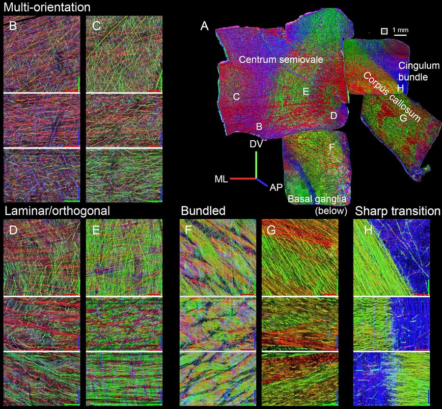
View on bioRxiv →Multi-orientation meshwork
Loosely packed axons traveling in many directions, forming an open, interwoven network.
Laminar/orthogonal
Alternating layers of near-orthogonal axons in a woven, lattice-like structure with periodic spacing.
Tight parallel bundles
Densely packed axons running in close parallel, optimized for direct inter-hemispheric transmission.
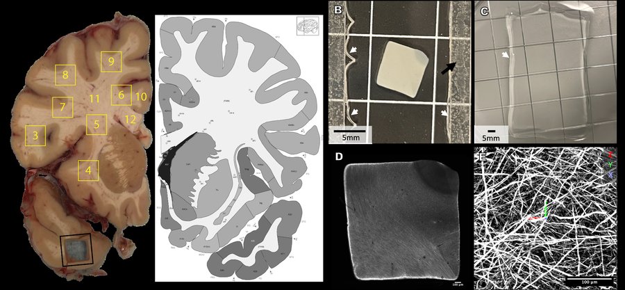
Human white matter
from slab to single axon.
The pipeline moves from post-mortem brain slabs to tissue punchouts, through expansion clearing, and into a custom ExA-SPIM lightsheet microscope — resolving individual axons at sub-micron voxels across centimeter-scale volumes.
More from the axonal
connectomics program.
A scalable and modular computational pipeline for axonal connectomics: automated tracing and assembly of axons across serial sections
A machine-learning pipeline for assembling axon traces across centimeter-scale serial sections, designed to scale toward whole-human-brain mesoscale connectivity mapping.
View on bioRxiv →Microscale visualization of cellular features in adult macaque visual cortex
Expansion microscopy combined with light-sheet imaging resolves fine-scale cellular features across large volumes of macaque visual cortex, using custom iSPIM microscopes and ~4× expansion protocols.
View on bioRxiv →EM connectomics &
the MICrONS program.
For many years the Reid lab was at the forefront of large-scale electron microscopy connectomics, producing some of the most complete synaptic-resolution circuit maps ever assembled. This work — now continued by colleagues at the Allen Institute — culminated in the MICrONS cubic millimeter dataset and a suite of papers in Nature in 2025.
Connectomics
The MICrONS program: functional connectomics of mouse visual cortex
High-throughput serial-section EM, co-registered with calcium imaging from the same neurons, produced a functional connectomics map of tens of thousands of neurons spanning primary visual cortex and higher visual areas. Data are freely available.
Explore EM Connectomics →Get in touch.
For research collaborations, data access, or general inquiries, please reach out.